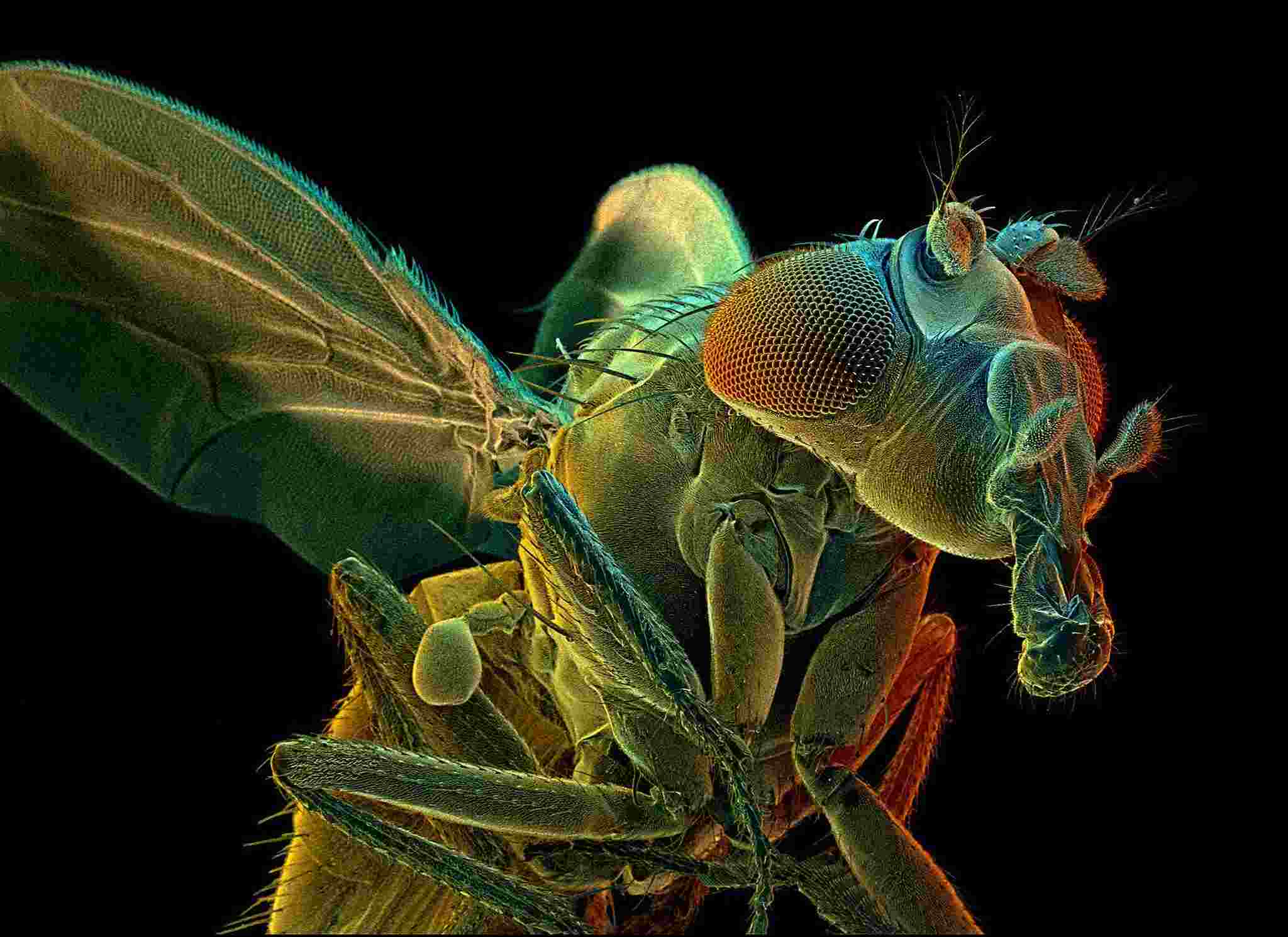
The brain of a fruit fly is approximately the size of a poppy seed, and it is just as simple to miss as it is to ignore the rest of the fly.
“I think most people don’t even consider the fly to have a brain,” he said. “But, of course, flies have quite a privileged existence.”
Even the simplest flies are capable of complex activities, including as traversing different environments, tussling with competitors, and serenading possible mates. They also have very sophisticated brains for their size, with around 100,000 neurons and tens of millions of connections, or synapses, between them.
Since 2014, a team of scientists at Janelia Research Center has been mapping these neurons and synapses in an attempt to produce a full wiring diagram, also known as a connectome, of the fruit fly brain in partnership with researchers at Google.
Although cutting-edge machine-learning algorithms are being used to speed up the process, the work is still time-consuming and costly. It is now in progress. Nonetheless, the information they have provided so far is breathtaking in its complexity, forming an atlas of tens of thousands of gnarled neurons in several critical parts of the fly brain.
And now, in a massive new research that will be published on Tuesday in the journal eLife, neuroscientists are starting to demonstrate what they can do with the information.
Dr. Jayaraman and his colleagues discovered scores of novel neuron types and found neural circuits that seem to assist flies in navigating their way around the environment by investigating the connectome of a tiny region of the fly brain — the central complex, which plays a key role in navigation. In the long run, the research might give valuable insight into how all sorts of animal brains, including our own, interpret a deluge of sensory input and transform it into appropriate behaviour.
It also serves as a proof of concept for the relatively new subject of contemporary connectomics, which was founded on the premise that creating precise maps of the brain’s wiring will provide scientific benefits in the long run.
It is “truly astonishing,” Dr. Clay Reid, a senior scientist at the Allen Institute for Brain Science in Seattle, said of the new work, which was published this week. “I believe that everybody who looks at it would agree that connectomics is a tool that we really need in neuroscience – period.”
The lowly roundworm, C. elegans, has the only full connectome known to exist in the animal realm. The study was initiated in the 1960s by the pioneering scientist Sydney Brenner, who would eventually go on to receive the Nobel Prize for his work. His tiny team worked on it for years, meticulously tracing each of the 302 neurons using colourful markers.
After establishing the Janelia Research Campus in 2006, Gerald Rubin, the campus’s founding director, set his eyes on the fruit fly as a research subject. I believe that flies have the simplest brains that are capable of engaging in interesting and complex conduct.”
A number of different teams at Janelia have undertaken fly connectome investigations over the years, but the study that culminated in the latest publication started in 2014 with the brain of a single five-day-old female fruit fly, which was used as a starting point.
In order to picture the fly brain, the researchers chopped it into slabs and then utilised a method known as focused-ion beam scanning electron microscopy to examine it layer by laborious layer. The microscope effectively functioned like a very small, very precise nail file, filing away an extremely thin layer of the brain, taking a photograph of the exposed tissue, and repeating the procedure until no more brain tissue was revealed.

